Oral and maxillofacial surgery in 3D.
Ramus is a workspace for dentists and surgeons: explain a treatment at the chair, plan it on real CT-derived anatomy, and learn each operation step by step.
Free for macOS and Windows, with two packages for Linux.
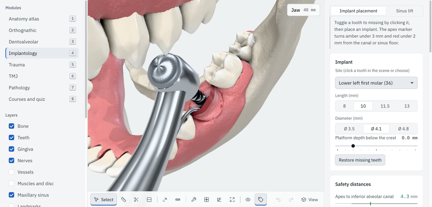
Show the patient what you will do.
Explain to patient has 23 topics, from a wisdom tooth to a Le Fort I. You talk and the model shows it.
-
A few cards in plain words.
Each topic has 3 to 5 cards. A card says one thing, such as “The gum is opened and folded aside”, and plays that part of the animation.
-
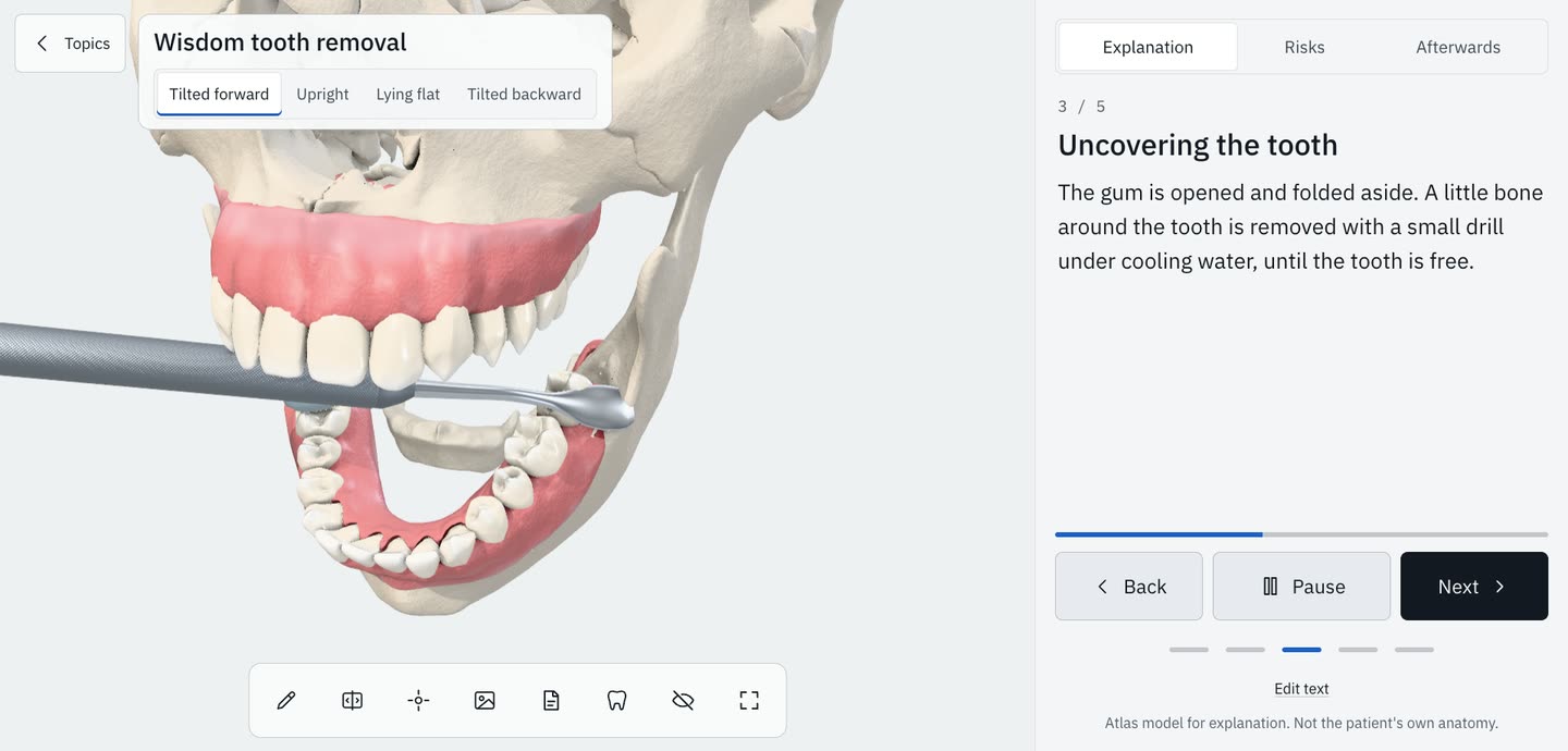
The risks, ticked as you talk.
Next and Back move through the cards. The risks are a list, and you tick each one when you have discussed it. Afterwards has the aftercare.
-
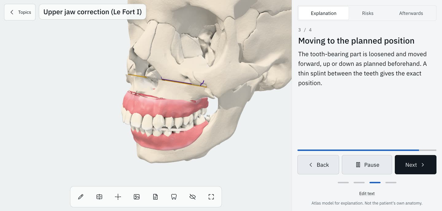
Draw on it. Print one page.
Draw on the 3D view with the pen, or place an arrow or a circle. The handout is one A4 page. Your drawing and the explanation are on it, and so are the risks you ticked.
Tap a tooth. The model follows.
The FDI chart and the 3D arch show the same teeth. Mark 36 as missing in one and it is gone in the other.
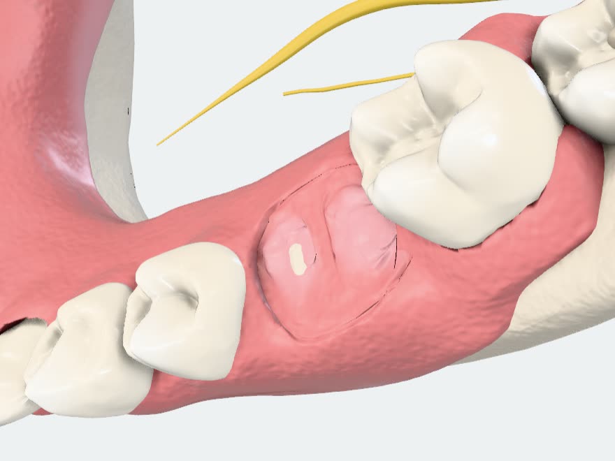
Cases stay on this device.
A case is stored on the device you work on and nowhere else. The chart and the plans belong to it.
One file, and a passcode if you want.
Export a case as one file when it has to move. Set a passcode and the stored cases are locked.
Plan with the nerve in view.
Import an STL, PLY, OBJ or GLB scan and align it to the model with three points.

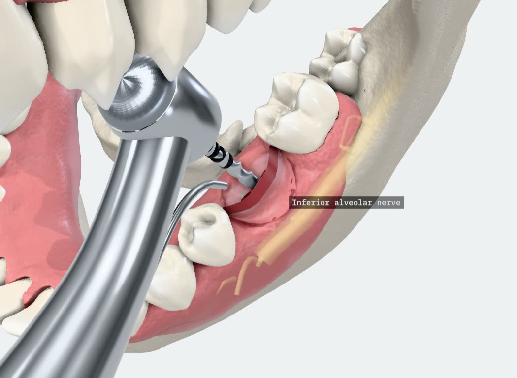
The distance, while you move the implant.
Ramus measures from the implant to the nerve canal and to the sinus floor as you place it. It checks the neighbouring teeth too. In this picture the apex is 4.3 mm from the canal.
The marker turns amber under 3 mm and red under 2 mm.
One tracing, six analyses.
SteinerRickettsMcNamaraJarabakWitsTweed
Trace a cephalogram on your own image and read it in any of the six. Orthognathic planning moves the jaw segments and lists each movement in millimetres.
34 operations, each on a timeline.
Every procedure is an animation you can stop anywhere. Drag the timeline back two seconds and watch the cut again.

Stop at any second.
A round bur opens a small window in the bone over the root apex.
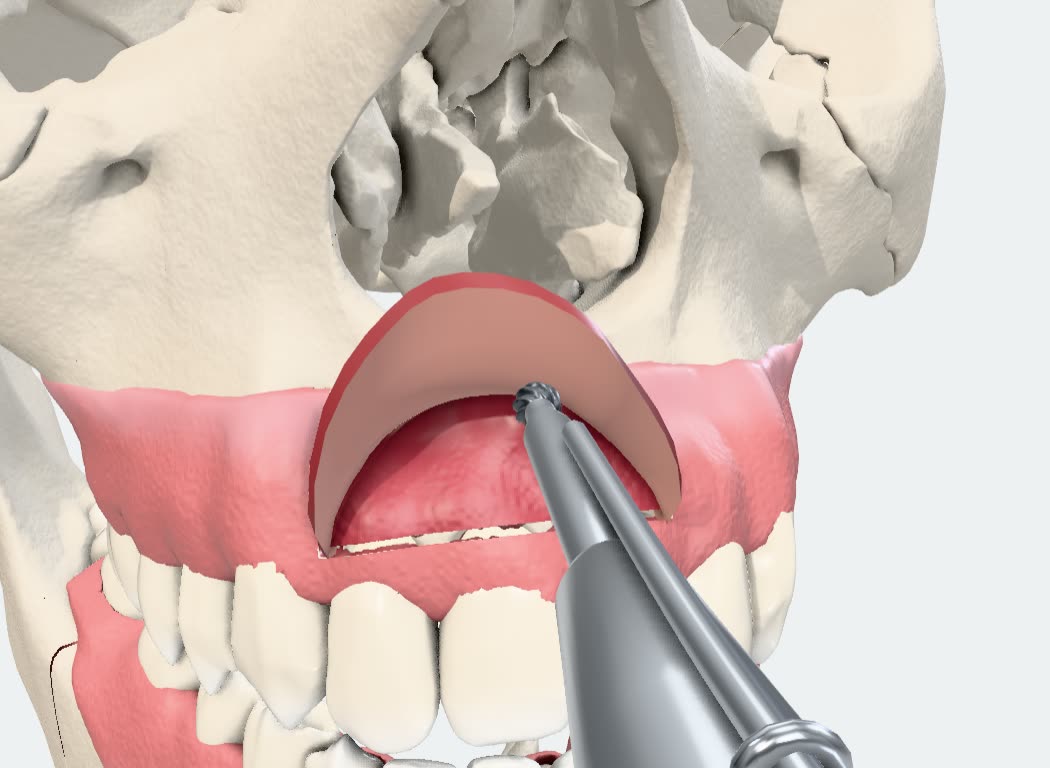
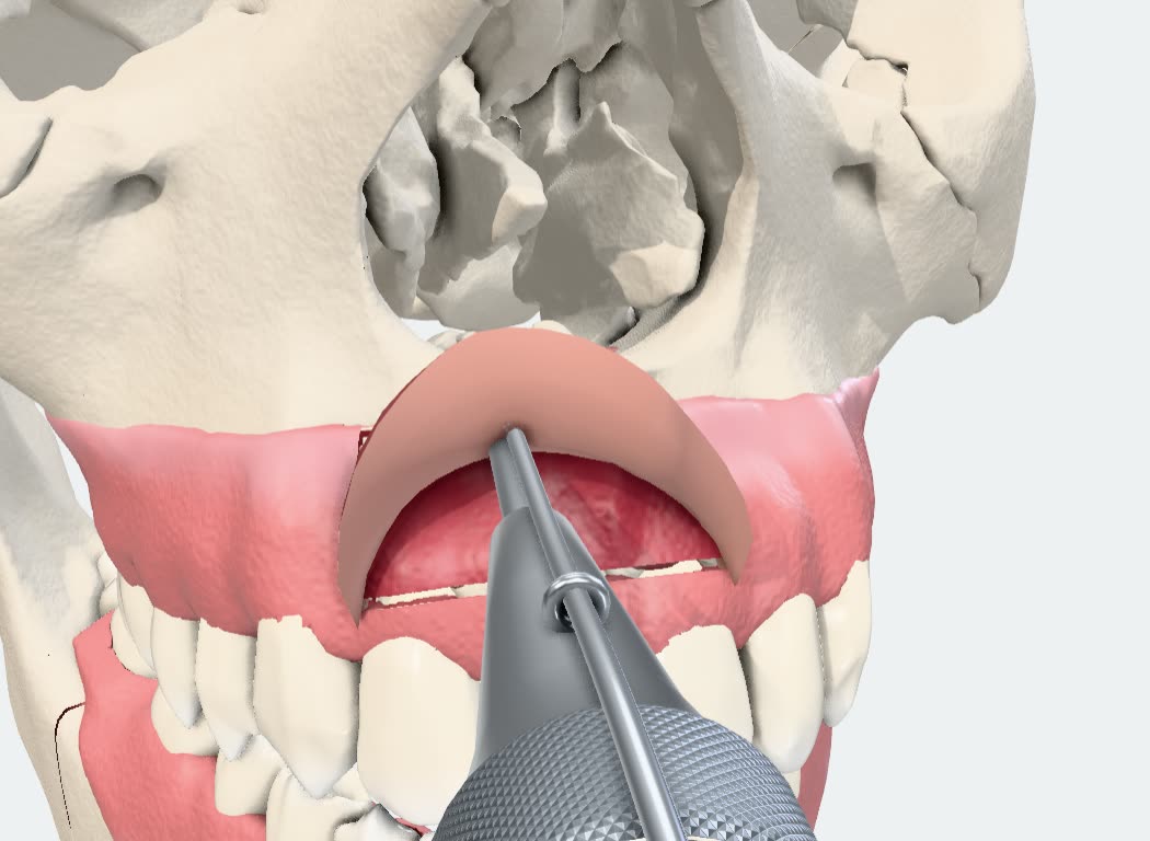
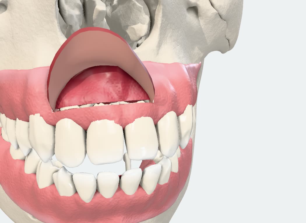
Apicoectomy of an upper incisor, 36 seconds. Three pictures of it are here; in Ramus the timeline is continuous.
Instruments touch what they cut.
A test measures every instrument against the bone and the teeth along its path. The bur meets the bone where the window opens, and the forceps close on the tooth.
In hands-on mode you hold the instrument yourself. Drag a saw and it cuts where you drag.
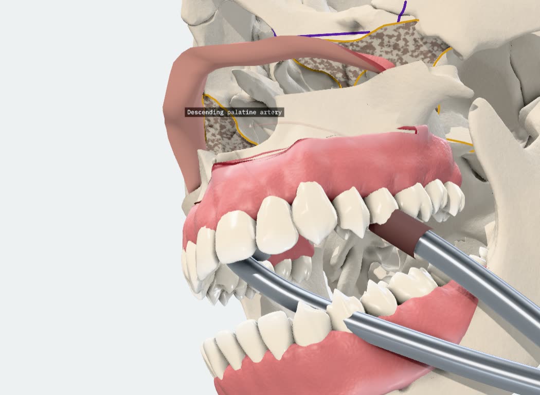
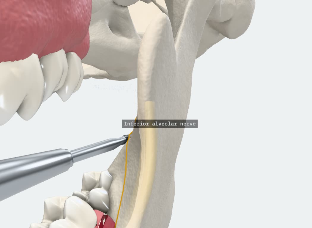
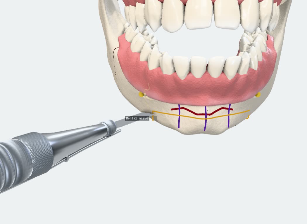
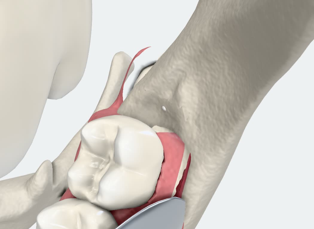
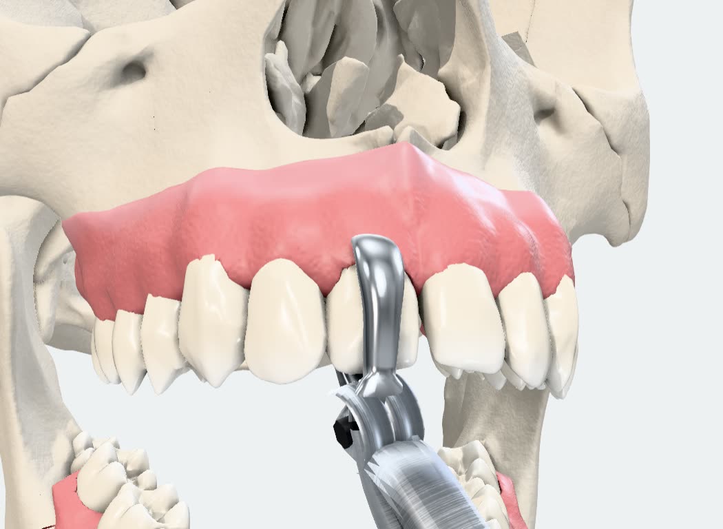
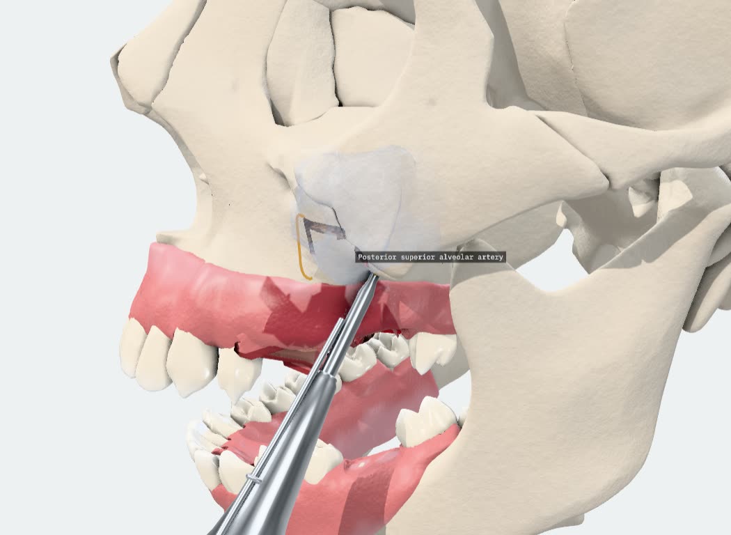
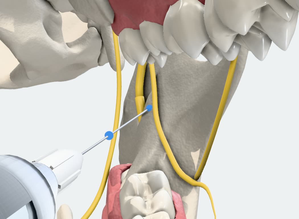
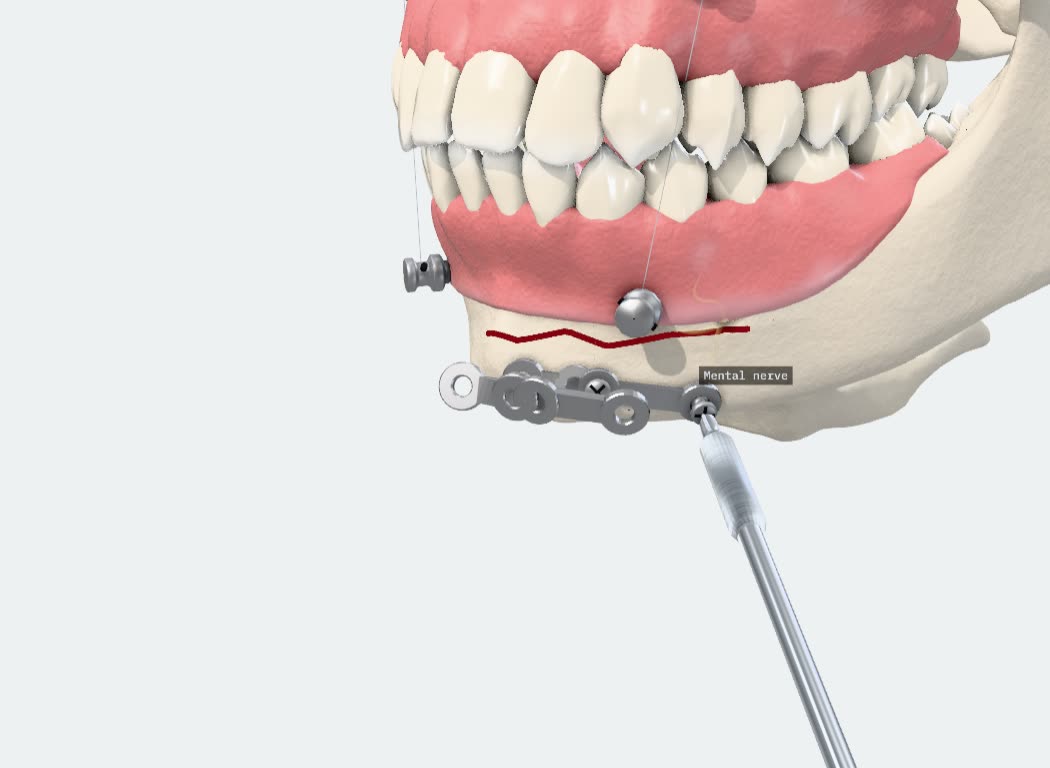
Also in the set: SARPE, implant placement, apicoectomy, arthrocentesis, cyst surgery, biopsy. Eleven of the 34 are nerve blocks.
86 cards. Each one names its source.
Every number on a card comes with its source. One search field finds the card.
- Local anaesthesia 5
- Nerve block atlas 11
- Antibiotics 4
- Analgesia 4
- Anticoagulation 6
- Medically compromised 6
- Emergencies 8
- Classifications 18
- Timelines 3
- Materials 6
- Post-operative instructions 9
- Differential diagnosis 5
- Cephalometric norms 1
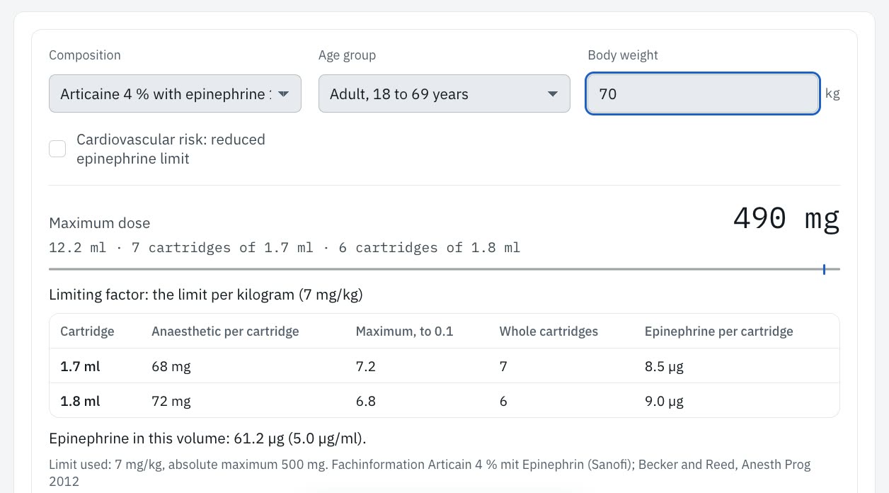
The maximum dose for this patient.
Pick the anaesthetic and the age group, then enter the weight. The calculator gives the dose in milligrams and in whole cartridges, and it says which limit it used.
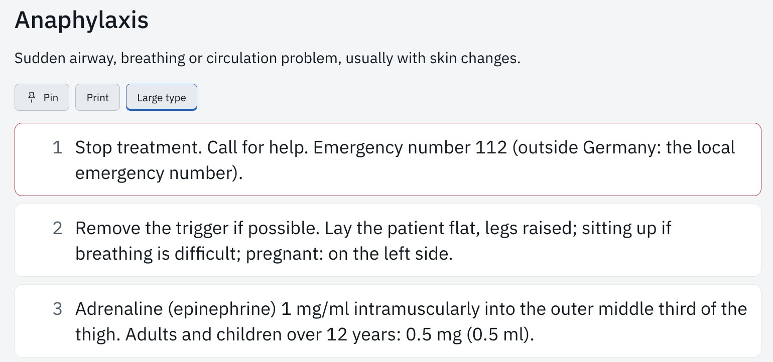
Emergencies in large type.
Seven emergency cards, each a short list of numbered steps. Anaphylaxis starts with “Stop treatment. Call for help.”

Nine courses. 307 questions.
Each course keeps your progress. A question you miss comes back after 10 minutes, then after 1, 3, 7 and 21 days.
- Anatomy38
- Local anaesthesia37
- Exodontia34
- Dentoalveolar surgery34
- Implantology33
- Trauma32
- Orthognathic surgery35
- Temporomandibular joint30
- Oral pathology34
Exam mode sets 10, 20 or 40 questions against a clock.
Patient data stays on the device.
Ramus has no account and no analytics. It works offline. A case leaves the device only when you export it.
Facts.
- Systems
- macOS on Apple silicon and Intel. Windows, 64-bit. Linux as AppImage and as deb package. Ramus also runs in a browser.
- Languages
- German and English.
- Anatomy
- Real CT-derived anatomy from BodyParts3D, © The Database Center for Life Science, licensed under CC Attribution-Share Alike 2.1 Japan. Nerve and vessel courses after Z-Anatomy, CC BY-SA 4.0. The jaw opens and closes.
- Explain
- 23 patient topics with drawing tools and a one-page A4 handout.
- Procedures
- 34 animated procedures, 11 of them nerve blocks.
- Reference
- 86 cards in 13 categories, each card naming its source.
- Learn
- 307 questions in 9 courses. Exam mode and spaced review.
- Scans
- STL, PLY, OBJ, GLB. Aligned with three points.
- Cephalometry
- Steiner, Ricketts, McNamara, Jarabak, Wits, Tweed.
- Data
- Stored on the device, with no account and no analytics. Works offline. A case exports as one file, and a passcode can lock it.
- Price
- Free.
- Use
- Teaching and planning aid. Not a medical device and not for diagnosis.
Download Ramus.
Free installers for the desktop.
The download buttons need JavaScript. With it off, the installers are on the GitHub releases page of 0xBakeer/ramus.
Ramus is a desktop app. Open this page on a Mac, a Windows PC or a Linux machine to install it.
-
Your system
macOS
Disk image
-
Your system
Windows
Installer, 64-bit
-
Your system
Linux
x86-64
Ramus is a teaching and planning aid. It is not a medical device and not for diagnosis.
likes
Leave a like.
Ramus is free. If it helped you explain a case or pass an exam, a like tells me so.
The coffee button opens GitHub Sponsors.